Leukemia and Malignant Blood Disorders Scientists
Dr. Versha Banerji
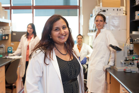
Dr. Versha Banerji is a Clinician Scientist at CancerCare Manitoba and Senior Scientist at the Research Institute in Oncology and Hematology. She is also a co-investigator on the Research Manitoba CLL Research Cluster.
Cancer cell metabolism is considered a hallmark of cancer development. Current treatment strategies are based on targeting DNA replication or signaling pathways to control cancer cell growth proliferation and survival. My laboratory firmly believes that targeting cancer cell metabolism is a novel approach in the fight against cancer. My research program is focused on evaluating the metabolism power house, the mitochondria, by creating bioenergetics profiles of cancer cells. We then use the readouts to determine how new drugs can alter the profiles and identify doses that may be safer in patients and better tolerated by patients for treatment of their cancers. Although my work has mainly focused on Chronic Lymphocytic Leukemia, this work has broad applicability to any cancer type. This platform serves to supplement traditional drug screening for novel agents. We also use a variety of molecular biology techniques in the laboratory and genetic loss of targets as validation for drug screens. We also investigate how cancer metabolism is linked with circadian rhythm. As a physician, I value the use of novel agents in clinical practice and how they affect my patients. Thus, my research program also evaluates the implementation of novel agents in clinical practice, the impact on treatment course of patients including side effects as well as the costs of new drug for health care delivery.
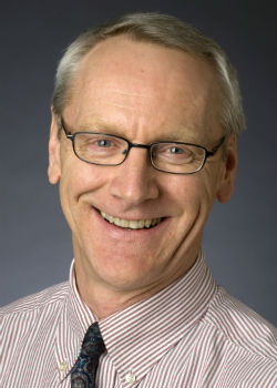
Spencer Gibson, PhD
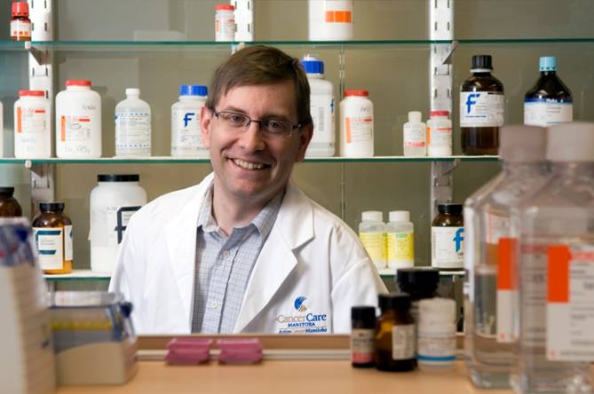
In maintaining integrity and homeostasis of multicellular organisms, the balance between cell death and survival is fundamentally important. When this balance is altered, diseases such as cancer occur. One protein important in the regulation of cell death is BNIP3 which is induced under low oxygen (hypoxia) conditions and is over expressed in solid tumours. This paradox of BNIP3 killing cancer cells while being over expressed in live cells within tumours is a focus of our research. Three explanations could account for these differences and act as a mechanism for cancer progression.
Cell survival is as important as cell death. The epidermal growth factor receptor (EGFR) is expressed at high levels in several cancers including breast cancer. We discovered that pretreatment of breast cancer cell lines with epidermal growth factor (EGF) effectively blocked drug and death receptor induced apoptosis. This protection from apoptosis is mediated by a serine threonine kinase (AKT) through up-regulation of the Bcl-2 anti-apoptotic family member Mcl-1. Besides breast cancer, we have found that a lipid, lysophosphatic acid (LPA) blocks apoptosis in chronic lymphocytic leukemia (CLL) cells using a similar mechanism. We are currently investigating the regulatory elements controlling Mcl-1 expression.
The goal of my research is to define the signal transduction pathways leading to cell death or survival. This will elucidate targets that could tip balance in favour of cell death and will be the foundation to establish clinical trials using molecular targeted therapies to increase effectiveness of chemotherapy in cancer.
Contact Information:
5008b-675 McDermot Ave
Winnipeg, Mb
R3E 0V9
Phone: 204-787-2051
Email: Dr Spencer Gibson PhD
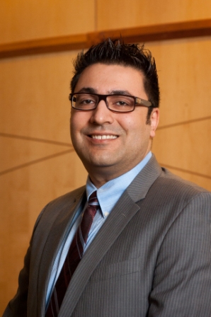
Haigh, Jody
Dr Haigh is an expert in mammalian functional genetics, has developed transgenic mouse models to investigate cell differentiation for leukemias, and has expertise in novel mouse embryonic and induced pluripotent stem cell to investigate hematopoietic development. He has 98 publications including recent papers in Blood, Nature Immunology, PLoS Genetics and PLoS One, Journal of Experimental Medicine, and multiple papers in EMBO Journal. His expertise in drug screening platforms will benefit the clinicians and scientists in CCMB, RIOH and at the University of Manitoba, allowing rapid identification of novel drugs or drug combinations to work more effectively in treating cancers.
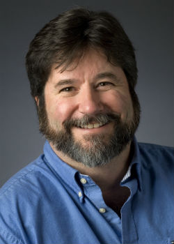
Geoff Hicks, PhD
Transformation
Mutations (from the ES cell library) transmitted to the germline will focus on genes known or suspected to be involved in tumour progression. Understanding the normal function of a gene in mammalian development is a powerful approach to understanding how the oncogene contributes to the respective cancer. The focus of the lab is on genes which are translocated in the development of human cancers; specifically, the TLS and EWS genes. While the translocations and the associated cancers for these genes are highly characterized, little is known about function of the genes themselves or how they contribute to tumour development. Our approach is to analyze developmental defects in mice that are either deficient for specific gene (and are otherwise genetically identical to wild-type mice). For example TLS is a gene that is translocated in many human soft tissue sarcomas and myelogenous leukemia. Functional analysis of mice that are homozygous for the TLS/FUS mutation has revealed TLS plays a critical role in the normal development of blood cells and in maintaining genomic stability. Using this approach, we have now discovered the how changes in TLS during cancer specifically prevent its normal role to limit cell proliferation and correct mutations in other genes -- both of which are hallmarks of cancer itself.
Contact information:
Regenerative Medicine Program
669 Basic Med Sci Bldg
745 Bannatyne Avenue
University of Manitoba
Winnipeg, Mb R3E 0J9
Phone: (204) 318-5287
Lab: (204) 318-5289
Email: Dr Geoff Hicks PhD
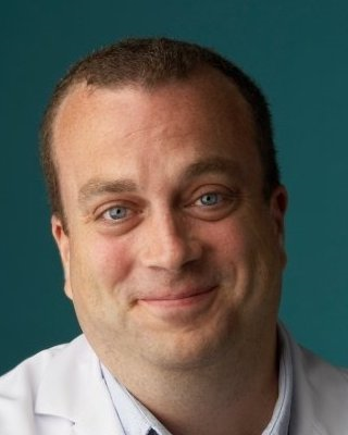
Dr James Johnston MB, BCh, FRCPC
My primary research interest is in chronic lymphocytic leukemia (CLL) and I am involved in a number of translational research programs related to this disease. These studies involve the epidemiology and basic science of CLL, in addition to clinical trials. To further these activities, we have developed the CLL Clinic at CancerCare Manitoba and the Manitoba CLL Tumour Bank, which is housed in the RIOH. Our epidemiological studies have demonstrated that the incidence of CLL is much higher than previously reported with elderly male patients having a particularly poor prognosis. Our ongoing laboratory studies are evaluating new therapies and prognostic markers in CLL and examining the effects of age and gender on the biology of this cancer.
Contact information:
Department of Medical Hematology and Oncology
2078-675 McDermot Ave
Winnipeg, Mb
R3E 0V9
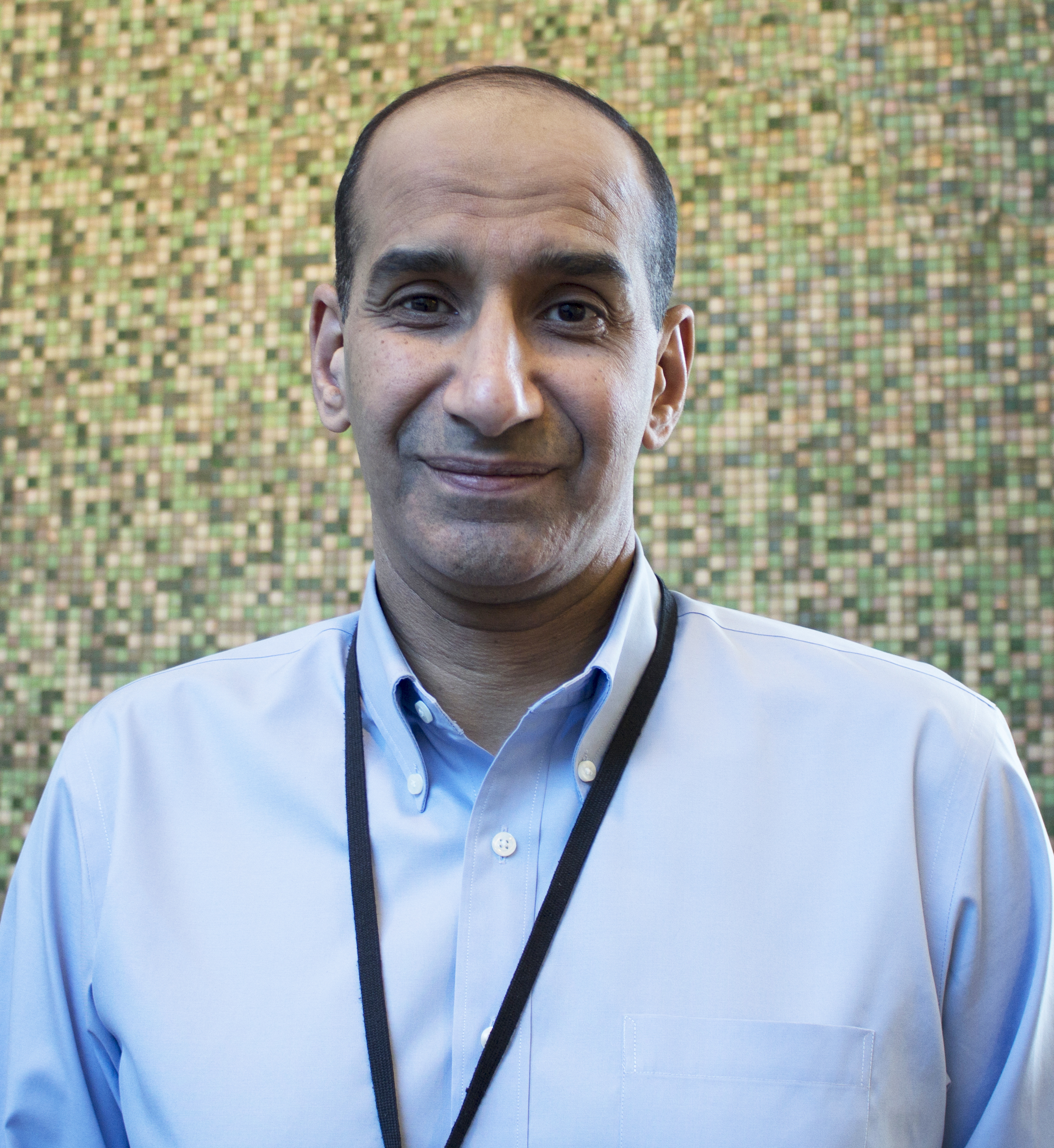
Dr Sachin Katyal, PhD
Modulating DNA Damage repair mechanisms to enhance brain tumour treatment success
DNA strand breaks occur on a daily basis in cells due to cell stress, environmental factors, oxidation and metabolism. Damaged DNA is resolved by dedicated DNA damage response (DDR) and repair mechanisms in order to preserve genomic integrity and cell function. The goal of conventional chemotherapeutic drugs and radiotherapy is to elicit DNA damage to overwhelm the tumour’s innate DDR and induce cell death. However, tumour cells have remarkable ability to respond to DNA damage, repair and adapt thus allowing survival and eventual drug resistance. It is predicted that >90% of all tumours incur at least one defect in the DNA damage response (DDR), thus tumour cell survival relies upon enhanced activity of other compensatory DNA repair pathways. The aggressive and deadly brain tumour, glioblastoma multiforme (GBM) shows a very high level of recurrence due to emergence of chemo/radio-resistant tumour cell populations; patients usually live about 1 yr from their date of diagnosis. We are identifying the “back-up” DNA repair pathways in these deadly brain tumours so that we can enhance the patients’ treatment success and their quality-of-life.
DNA repair pathways are guardians of the cellular genome.
Every individual human cell is estimated to incur tens of thousands of DNA strand breaks due to environmental stress, oxidation, metabolic function and DNA decay. To preserve genomic integrity, these breaks are resolved by dedicated DNA damage repair (DDR) pathways that ensure faithful transmission of genomes in dividing cells to ensure proper cell function and survival. There are two classes of DNA strand breaks, double-strand breaks (DSBs) and DNA single-strand breaks (SSBs), which are resolved by specific repair pathways, DSBR and SSBR. The inability to properly process and repair SSBs can interfere with the DNA replication and transcriptional machinery resulting in persistent SSBs, formation of the particularly genotoxic DNA double-stranded break (DSB) lesion and aberrant gene expression resulting in a variety of cellular pathology, including: senescence, cancer and apoptosis. It is known that the diverse mechanisms involving cell cycle regulation, DDR pathways, cellular metabolism, and cell death act in concert in response to DNA damage. As such, cellular life and death decisions are balanced by these mechanisms as defective DDR in proliferating cells, including neuroprogenitors, can lead to cancer, while defective neuronal DDR can lead to neurodegeneration.
Anti-tumorigenic agents overwhelm cellular DNA repair responses with lethal levels of genotoxicity.
The objective of common front-line radiation and chemotherapeutic strategies used in the treatment of brain tumours is to induce DNA breaks so as to overwhelm the cellular DNA repair machinery thus promoting genomic damage and tumour cell death. However, as the intrinsic cellular DNA repair process counteracts the therapeutic efficacy of this strategy, high radiation and drug doses are required which result in harmful neural and systemic side effects. My research seeks to identify ways to dysregulate cellular DNA repair pathways in tumours and improve therapeutic success. In this regard, DNA damage repair pathways are an ideal clinical target as we can specifically kill cancer cells by lowering the radio- and chemotherapeutic threshold of tumour cell genotoxicity by inhibiting redundant DNA repair pathways.
My research uses advanced molecular, biochemical and genetic techniques to gain insight into the biology of mammalian DNA strand break repair pathways. My goal is to identify ways to manipulate these pathways to develop novel treatment strategies in the clinical management of cancer. We are seeking a very highly motivated postdoctoral fellows (with a strong history of previous success) to join our team.
Contact information:
675 McDermot Ave
Winnipeg, Mb R3E 0V9
ph.: 204-787-2765, fax: 204-787-2190
Email: Dr Sachin Katyal PhD
Dr. Rami Kotb
Dr. Sabine Mai
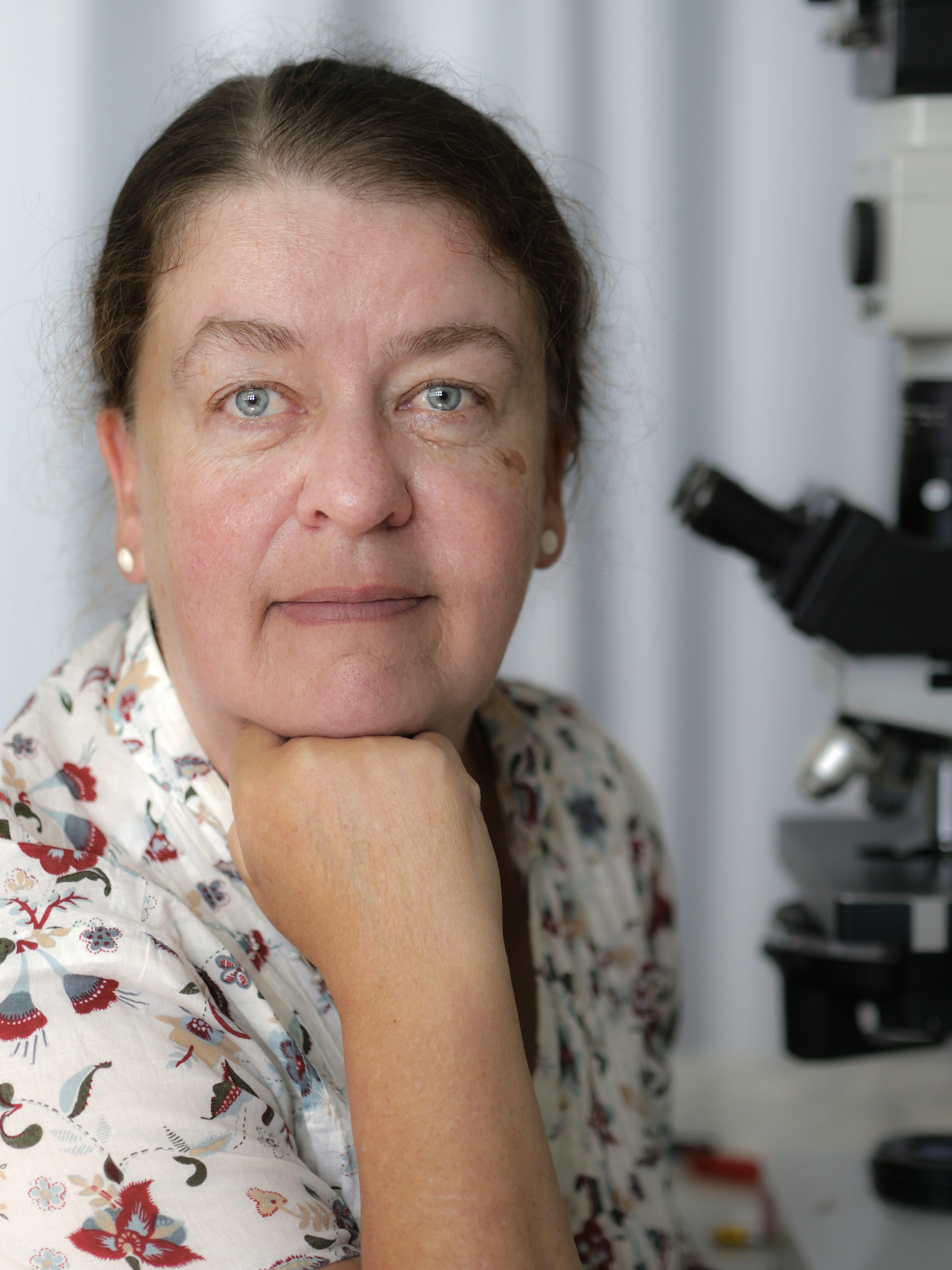
Dr. Mai was the first to show that the oncogene c-MYC drives the onset of genomic instability and nuclear genome remodeling, which she identified as a characteristic feature of cancer cells. This concept was recently recognized as a critical advance in molecular medicine and featured in the Cold Spring Harbour Perspectives in Medicine book “Myc and the pathways to cancer”. Dr. Mai was first to demonstrate telomere-dependent nuclear genome remodeling in cancer that serves as a mechanism of tumor initiation and progression, and also as a structural biomarker for cancer aggressiveness. To measure cancer genome remodeling, she applied quantitative 3D nuclear telomere imaging and analysis tools in translational research studies to multiple cancer sites including Hodgkin’s lymphoma and multiple myeloma. Her work showed that 3D nuclear telomere analysis is a powerful tool to stratify patients into distinct groups of stable and aggressive disease, which will, upon successful translation to the clinic, enable personalized cancer treatment decisions. This novel approach of 3D nuclear re-organization as a structural biomarker in cancer provides the first evidence that 3D nuclear telomere organization can be used as a hallmark of clinical decision-making.
Dr. Mai created the Genomic Centre for Cancer Research and Diagnosis (**GCCRD** link) at U Manitoba, a world-leading imaging facility with four grants from the Canada Foundation for Innovation (over $13M). The GCCRD features the first super resolution imaging facility in North America. Dr. Mai was first to study the 3D spatial organization of the cancer cell genome by super resolution microscopy, allowing for unprecedented insights into the cancer cell genome.
Over her career, Dr. Mai has trained over 1500 national and international students, postdoctoral fellows, scientists and clinicians, and has conducted advanced imaging courses in other countries including Thailand which helped in setting up 34 new imaging facilities there. Over the past 5 years, Dr. Mai has given 46 invited lectures, organized an international Imaging Symposium (2014), co-organized World Cancer 2016 in London (UK), and chaired sessions in this and other conferences. In 2015, Dr. Mai was recognized as one of the “Top 100, Canada’s Most Powerful Women” in the category of trailblazer and trendsetter to honor her engagement in research and training, and her work in translating her research from the bench to the bedside.
Contact information:
6046-675 McDermot Ave
Winnipeg, Mb R3E 0V9
(204) 787-2135
Email: Dr Sabine Mai PhD
Kristjan Paulson, MD
Dr. Paulson’s interests are focused in the areas of clinical effectiveness and health services research in acute leukaemia and blood and marrow transplantation (BMT). He is the Scientific Director of the Canadian Blood and Marrow Transplant Group Registry, a prospectively collected database with detailed clinical data on over 10,000 Canadians who have undergone BMT. He also is the Registry Lead for the Canadian National Transplant Research Program, a CIHR funded transplant research network, and has conducted multiple studies reviewing disparities in access to transplant and other complicated health services interventions.
Contact information
ON2078-675 McDermot Ave
Winnipeg, Mb
R3E0V9
For more information please contact:
CancerCare Manitoba
Phone: (204) 787-2197
Toll-free: 1-866-561-1026
Copyright © 2000-2019 CancerCare Manitoba.
All Rights Reserved.
 Accreditation Canada")